Key Findings

Brain neurons involved in Pavlovian learning patterns (i.e., learning in which a biologically potent stimulus [e.g. food] is paired with a previously neutral stimulus [e.g. a sound or tone]) shift their behaviors during this process, going from randomized to more synchronized patterns, as the behavior is learned. Understanding this memory mechanism could support future therapies for memory-related diseases.
About the Co-Author
Xuanmao Chen, Associate Professor of Molecular, Cellular, and Biomedical Sciences
Contact information: Xuanmao.Chen@unh.edu, 603-862-4542, Chen Lab website
This research first published in the Journal of the Federation of American Societies for Experimental Biology.
Researchers: Y. Zhou, L. Qiu and X. Chen
In an examination of the brain’s hippocampus (the area responsible for long-term memory formation), COLSA researchers discovered that the neurons involved in so-called Pavlovian conditioning (also known as respondent or classical conditioning) shift their behaviors during this learning procedure, becoming more synchronized when a memory is being formed.
Their finding improves understanding of memory mechanisms and offers clues for the development of future therapies for memory-related diseases and disorders like dementia, autism and post-traumatic stress disorder (PTSD). The researchers published their study findings in an issue of the Journal of the Federation of American Societies for Experimental Biology.
“There are tens of millions of neurons in the hippocampus but only a small fraction of them are involved in this learning process,” says Xuanmao (Mao) Chen, co-author and an assistant professor in UNH's molecular, cellular, and biomedical sciences department. “Before engaging in Pavlovian conditioning, these neurons are highly active, almost chaotic, without much coordination with each other, but during memory formation they change their pattern from random to synchronized, likely forging new connecting circuits in the brain to bridge two unrelated events.”
"Before engaging in Pavlovian conditioning, these neurons are highly active, almost chaotic, without much coordination with each other, but during memory formation they change their pattern from random to synchronized, likely forging new connecting circuits in the brain to bridge two unrelated events."
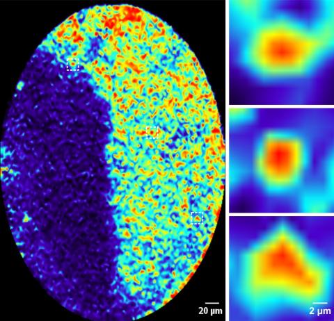
On the, the enlarged image shows many hippocampal neurons, most of which are silent (blue) and only a few are active. On the right are close-up images of three highly active neurons, or memory cells, which become synchronized after memory formation. Images modified from Zhou et al., 2020 FASEB Journal.
In the study, the researchers looked at Pavlovian learning patterns in mice. In the beginning, before any repetitive learning exercises, the mice did not know what to expect; using special imaging with an endomicroscope, the researchers could see that neural activity was disorderly. But after repeating different tasks associated with a conditional stimulus, like a tone or bell, the mice began to recognize the pattern and the highly active neurons became more synchronized. The researchers hypothesize that without forming synchronization, animals cannot form or retrieve this type of memory.
This work was supported by the National Institutes of Health and the Cole Neuroscience and Behavioral Faculty Research Awards. Co-authors include Y. Zhou, L. Qiu and X. Chen.
Related Links
Contact
